Fundamentals of biomedical imaging
PHYS-438
Media
Media
Welcome !
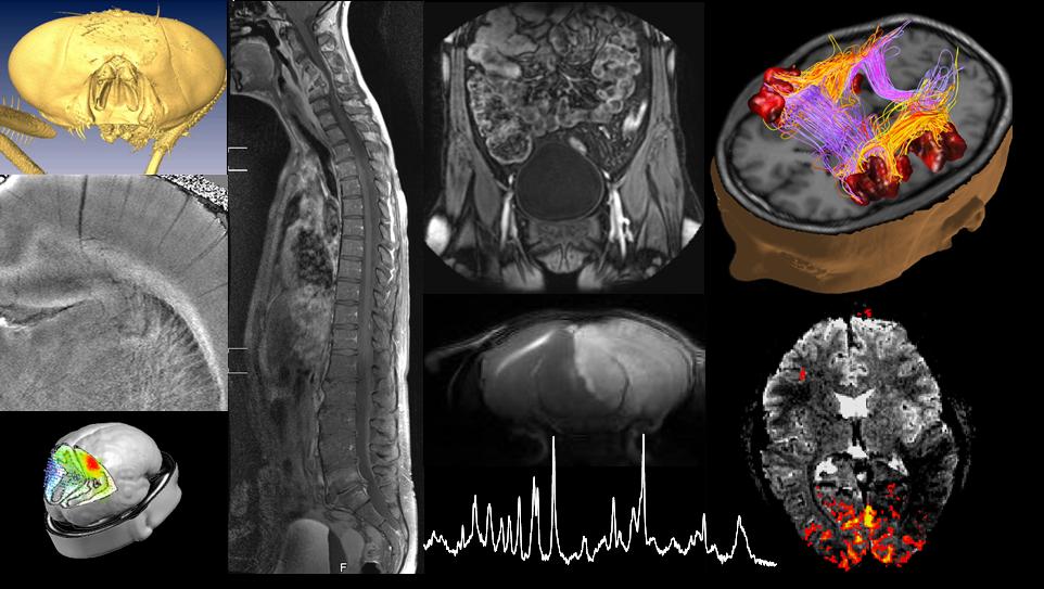
This course covers the physical principles of major in vivo bio-imaging modalities, i.e., ultrasound, computed tomography, emission computed tomography, positron emission tomography and magnetic resonance imaging. We will show how existing physical principles transcend into bio-imaging and establish an important link into life sciences, illustrating the contributions physics can make to life sciences.
In the lectures, practical examples will be shown to illustrate the respective imaging modality, its use, premise and limitations, and biological safety will be touched upon.
The student will develop a good understanding of the mechanisms leading to tissue contrast of the bio-imaging modalities covered in this course, including the inner workings of the scanner and how they define the range of possible biomedical applications.
Based on this course, the student will be able to judge which imaging modality is adequate for specific life science needs and to understand the limits and promises of each modality.
The course was live broadcast in 2014 (and partially in 2015), using google+ hangout on air, the videos are available also on our dedicated youtube channel, FundBioImag. The links to the recorded courses are provided on this site (in the content description of the respective week).
In addition, the course is available in the MOOC format (recording in the studio) in two parts:
Part I: https://courseware.epfl.ch/courses/course-v1:EPFL+PHYS438+2019_1/about
Part II: https://courseware.epfl.ch/courses/course-v1:EPFL+PHYS438a+2019_1/about
- You can arrange for a meeting, outside the exercis... (Text and media area)
- Exam 2025 - Info (Page)
- News forum (Forum)
- (Text and media area)
- Discussion forum (Forum)
1: Introduction (pdf)
- Course organization
- Bio-Imaging in Life sciences
- Essential elements of bio-imaging (Webb Ch. 5.3 & 5.4)*
- Lab visit
*indicates approximate correspondence with the book of Andrew Webb, but course may be more extensive or more qualitative. It is not recommended to use the book other than as additional support.
|
2: Ultrasound and Introduction to x-rays (pdf)
- Introduction to ultrasound imaging (Ch. 3.8, 3.10)
- Ionizing radiation
- Generation of ionizing radiation (Ch. 1.2)
|
3: Interaction of x-rays with tissue (pdf)
- Linear attenuation coefficient (1.4)
- Interaction of photons with matter (1.5)
Rayleigh scattering
Compton Scattering
Photoelectric effect
Pair production
- Effective atomic number Zeff (1.7)
- Radioprotection - a short primer (1.14)
Following this week you
|
4: From x-ray to image: Computed tomography (pdf)
Exercice session in CM 221 today
- Experimental principle (1.10)
Beam hardening effects
Sensitivity and resolution considerations
- Intensity projection (1.11)
Radon Transform
Projection reconstruction (App. B)
Sinogram
- Central slice theorem
|
5: Emission computed tomography (pdf)
- Experimental principle (2.1)
Tracers
- Resolution issues: collimators (2.7)
- Attenuation correction (2.9)
- x-ray detection - scintillation followed by amplification (2.7)
- Scatter correction (2.9)
|
6: Positron Emission Tomography (2.11) (pdf)
- Coincidence detection - electronic collimation
- Scatter and random correction
- Attenuation correction
- Factors affecting resolution
|
7: Tracer kinetics (pdf)
- Compartment models
- Fick's principle
- One-tissue compartment model
- Modeling tracer uptake
- deoxyglucose uptate
|
8: Introduction to magnetic resonance (4.1, 4.2, 4.4) (pdf)
- The MR scanner
- Magnetism of the nucleus (spin)
- The basic equation of motion of MR: precession
- Rotating frame of reference
After this course you
|


9: Relaxation of nuclear magnetization (4.2) (pdf)
- Detection (and excitation) of the MR signal
- Rotating frame revisited
- Resonance: effective RF field and flip angle
- Relaxation
- T1 and T2
- Bloch equations
- Relaxation basics: free induction decay (FID)
|
10: NMR spectroscopy (4.10) (pdf)
| 1. can calculate the effect of multiple RF pulses on longitudinal magnetization 2. know the definition of Ernst angle 3. Understand the two basic mechanisms by which electrons influence the precession frequency of nuclear magnetization 4. Know the definition of chemical shift 5. Know how and under what molecular conditions NMR spectroscopy can provide non-invasive biochemical information |
11: Echo formation and spatial encoding (4.3) (pdf)
- The 3rd magnetic field: Gradient
- Spatial encoding
- Gradient dephasing and rephasing
Gradient echo sequence
k-space
|
- Spatial encoding - additional info/alternate view (File)
- The above in pps (File)
- Problem set #11 (File)
- Solutions #11 (File)
12: MRI contrast mechanisms (pdf)
- T2*-weighted contrast: BOLD fMRI (4.11)
- Hahn spin echo (4.2)
- T1-, T2- and proton-density weighted MRI
- MRI contrast agents (4.7)
|
13: Magnetic resonance: Advanced contrast mechanisms & Overview of imaging modalities (pdf)
- Effect of flow
- Diffusion-weighted MRI (4.9)
- Anisotropic diffusion
Limitations
- Comparison by examples
|